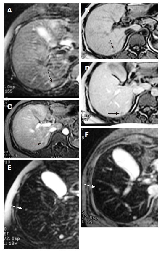
Figure 2.

Liver metastasis from mammary adenocarcinoma. A, B: Only one lesion (black arrow) was found on the pre-contrast T2WI and T1WI GRE images; C, D: Only one lesion (black arrow) was found on dynamic T1WI GRE images obtained after administration of Gd-DTPA; E, F: An additional small metastasis (0.3 mm, white arrow) was detected on T2WI and T2*WI images during the accumulation phase.
