
Figure 3.
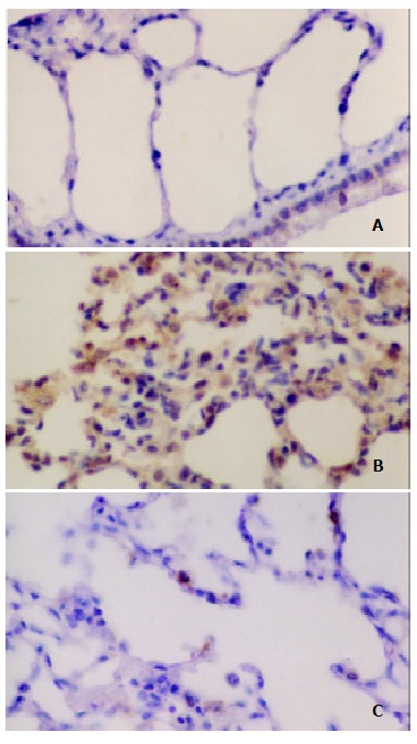
Immunohistochemical analysis of NT in the lung after IIR with pretreatment of AG in rats. SP stain × 400. A. No positive signal was found in the lung in sham group; B. Intense positive NT staining was found in the IR group; C. Positive NT staining decreased in the IR + AG group.