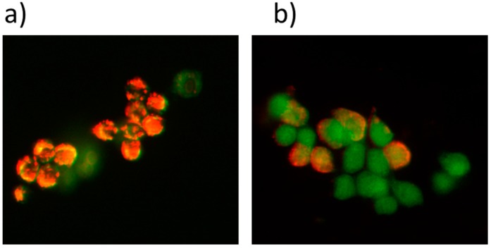
Figure 6.
Effect of LDH nanoparticles on mitochondrial membrane potential in HT-29 cells. (a) Control cells with LDH–ICG nanoparticles show most cells had a strong J-aggregation (red) in the absence of light irradiation; (b) drug-loaded LDH nanoparticles show a majority of these cells emitted green fluorescence, due to low mitochondrial membrane potential after treatment with LDH–NH2–ICG under light irradiation (magnification of both images at 40×).

