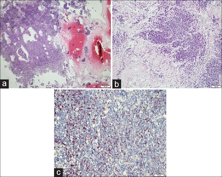
Figure 2.

Histological examination of the resected hemorrhagic mass. (a) Necrotic and hemorrhagic tumor tissue consistent with medulloblastoma (H and E, ×40). (b) Highly cellular tumor tissue was composed of primitive neuroepithelial cells; necrosis was present (H and E, ×100). (c) Numerous tumor cell nuclei stained positive with the Ki-67 monoclonal antibody (Anti-Ki-67, ×400)
