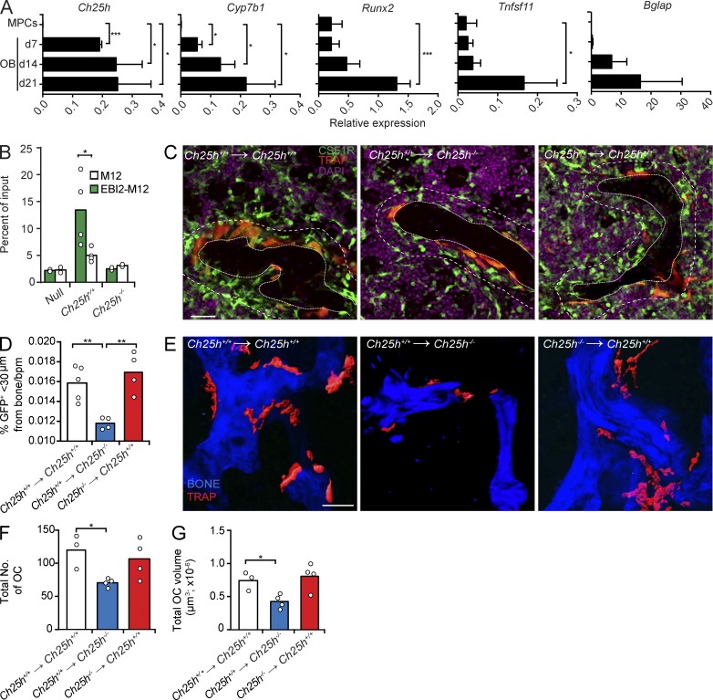
Figure 8.
CH25H expressed in radiation-resistant cells promotes OCP positioning near bone and OC differentiation. (A) Ch25h, Cyp7b1, Runx2, Tnfsf11, and Bglap mRNA expression in MPCs and in OBs differentiated in vitro for 7, 14, and 21 d. Bars indicate mean ± SD of triplicate measures. (B) Migration of M12 cells overexpressing EBI2 or control cells toward OB culture supernatants. Data are representative of four independent experiments. (C) Analysis of CSF1R+ cells and TRAP+ OCs by confocal microscopy in situ. Dashed lines delineate areas <30 µm distal from bone, and dotted lines delineate trabecular bones. (left and middle) Lethally irradiated Ch25h+/+ and Ch25h−/− mice were reconstituted with Ch25h+/+ CSF1R-GFP;TRAPRed BM cells. (right) Lethally irradiated Ch25h+/+ mice were reconstituted with Ch25h−/− CSF1R-GFP;TRAPRed BM cells. (D) Quantification of CSF1R+ cell proximity to bone surfaces: total number of CSF1R+ cells that are <30 µm distal from bone was divided by total number of CSF1R+ cells in the field of view and multiplied by 100 (%). The percentage of bone-proximal CSF1R+ cells was subsequently divided by the trabecular bone perimeter (bpm). Data are representative of at least four independent experiments. (E) Two-photon microscopy of femurs recovered from the BM chimeras described in C. (C and E) Bars, 50 µm. (F and G) OC total number (F) and total volume (G) in femurs from BM chimeras described in C. (A, F, and G) Data are representative of at least three independent experiments. (B, D, F, and G) Bars indicate mean, and circles indicate individual experiments. *, P < 0.05; **, P < 0.01; ***, P < 0.001 by unpaired Student’s t test.

