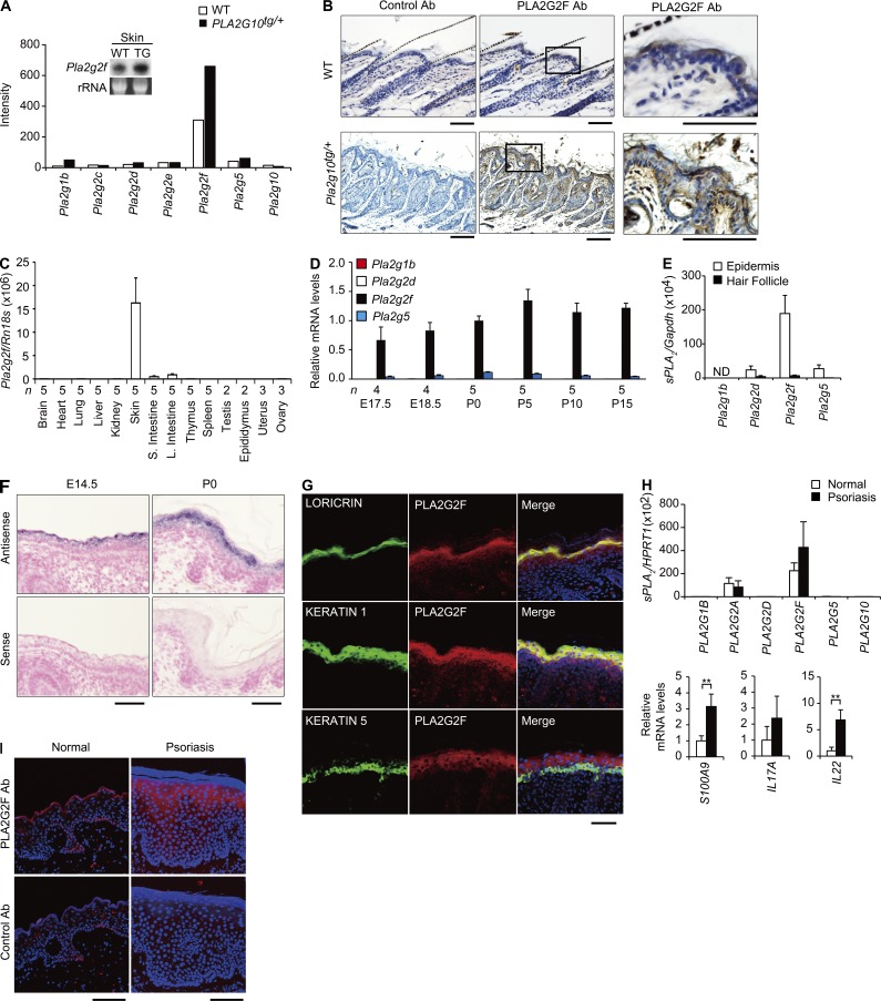
Figure 1.
PLA2G2F is expressed in the suprabasal epidermis. (A) Expression of sPLA2s in PLA2G10tg/+ (TG) and WT skins at P25, as evaluated by microarray. (inset) Northern blotting of Pla2g2f, with ribosomal RNA (rRNA) in an agarose gel with ethidium bromide as a control. (B) Immunohistochemistry of PLA2G2F in PLA2G10tg/+ and WT skins at P25 (bars, 100 µm). Boxes (middle) are magnified on the right. (C) Quantitative RT-PCR of Pla2g2f in various tissues of 8-wk-old C57BL/6 mice. (D) Quantitative RT-PCR of sPLA2s in developmental skins of C57BL/6 mice, with expression of Pla2g2f at P0 as 1. (E) Microdissection followed by quantitative RT-PCR of sPLA2s in the epidermis (n = 5) and hair follicles (n = 6) of C57BL/6 mice at P8. (F) In situ hybridization of C57BL/6 skin with an antisense or sense probe for Pla2g2f (bar, 50 µm). (G) Confocal immunofluorescence microscopy of PLA2G2F (red), keratinocyte markers (green) and their merged images (yellow) in newborn C57BL/6 skin (bar, 100 µm). (H) Quantitative RT-PCR of sPLA2s and psoriasis markers in normal and psoriatic human skins, with expression in normal skin as 1 (n = 7). (I) Immunohistochemistry of PLA2G2F (red) in human normal and psoriatic skins, with DAPI counterstaining (blue; bar, 100 µm). Data are from one experiment (A, E, and H) or are representative of two experiments (C, D, and A [inset]; mean ± SEM; *, P < 0.05; **, P < 0.01). Images are representative of two experiments (B, F, G, and I). ND, not detected.

