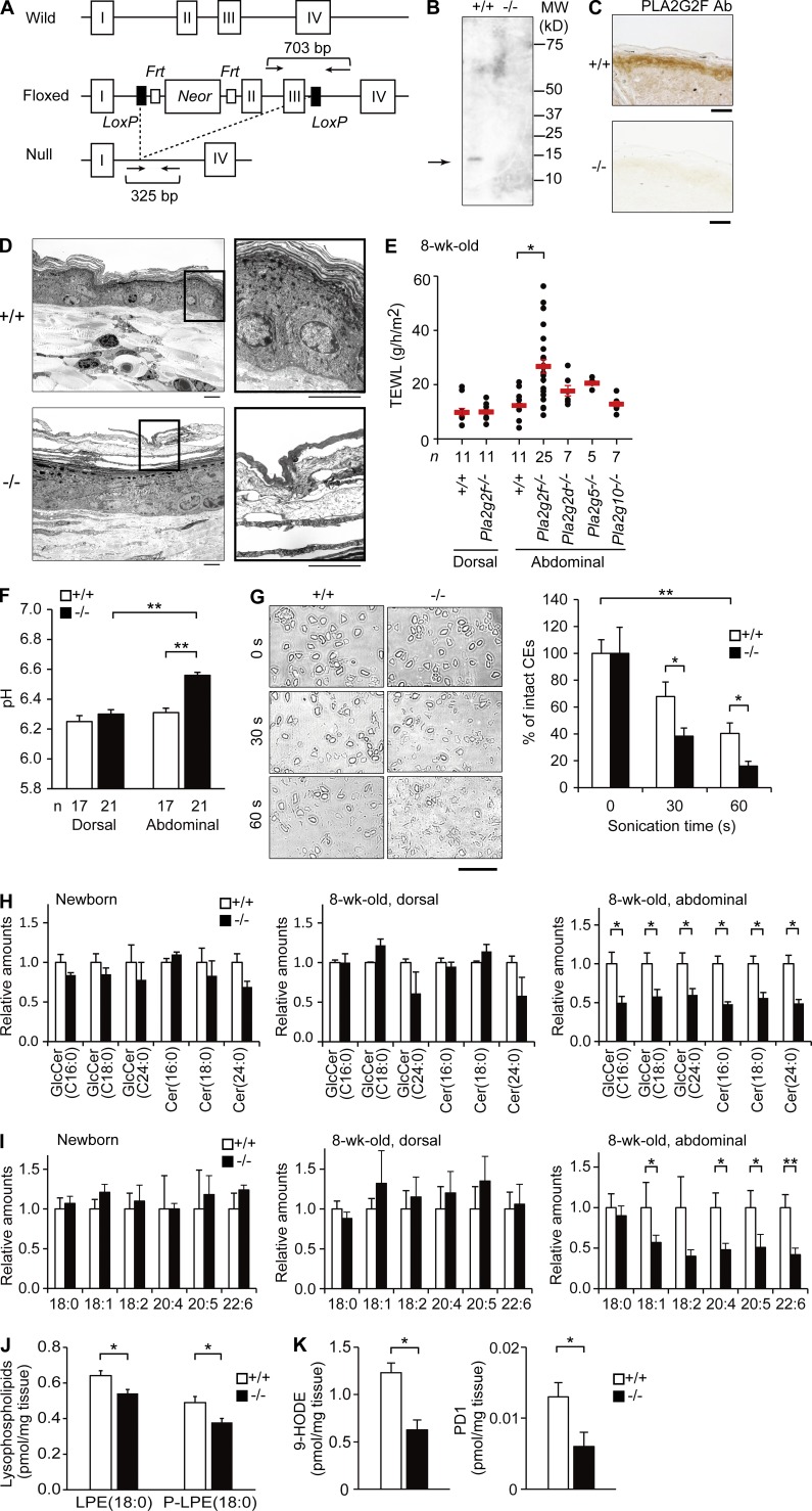
Figure 3.
SC abnormalities in Pla2g2f−/− mice. (A) Generation of Pla2g2f−/− mice. The Pla2g2f-targeting vector was constructed with the Neor gene that was inserted between exons 1 and 2 of the Pla2g2f gene. After mating with CAG-Cretg/+ mice, the exons 2 and 3 plus the Neor casette were removed to generate Pla2g2f-null mice. Arrows indicate primer positions for genotyping. (B) Immunoblotting of PLA2G2F protein in the SC of adult Pla2g2f+/+ and Pla2g2f−/− mice. (C) Immunohistochemistry of PLA2G2F in Pla2g2f+/+ and Pla2g2f−/− skins at P0 (bars, 100 µm). (D) Transmission electron microscopy of abdominal skins of Pla2g2f+/+ and Pla2g2f−/− mice (bar, 5 µm). Boxes are magnified on the right. (E) TEWL of dorsal and abdominal skins from various sPLA2-null or WT mice. (F) pH of dorsal and abdominal skins from Pla2g2f+/+ and Pla2g2f−/− mice. (G) Microscopic images of corneocytes (CEs) after sonication for the indicated periods (bar, 50 µm). (right) Percentages of intact cells (n = 7). (H and I) ESI-MS of ceramides (n = 6; H) and fatty acids (n = 6; I) in newborn and adult (dorsal and abdominal) Pla2g2f+/+ and Pla2g2f−/− skins, the values for WT skin being 1. (J and K) ESI-MS of LPE and P-LPE (J) or 9-HODE and PD1 (K) in abdominal Pla2g2f+/+ and Pla2g2f−/− skins (n = 6). Data are representative from two experiments (G–K) or compiled from three experiments (E and F; mean ± SEM; *, P < 0.05; **, P < 0.01). Representative images of one or two experiments are shown (B, C, D, and G). Cer, ceramide; GlcCer, glucosylceramide.

