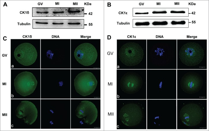
Figure 1.

Expression and subcellular localization of CK1δ and CK1ϵ during mouse oocyte meiotic maturation. (A, B) Expression of CK1δ (A) and CK1ϵ (B) were measured by western blotting. Samples (100 oocytes) were collected after oocytes had been cultured for 0, 8h and 12h, corresponding to GV, MI and MII stages respectively. Samples were immunostained for CK1δ or CK1ϵ, α-tubulin was stained as loading control. (C, D) Confocal microscopy showing the subcellular localization of CK1δ (A, green) and CK1ϵ (B, green) in mouse oocytes at GV, MI and MII stages. Oocytes at various stages were fixed and stained with anti-CK1δ or anti-CK1ϵ antibody, DNA (blue) was counterstained with Hoechst 33342. Bar=20 μm.
