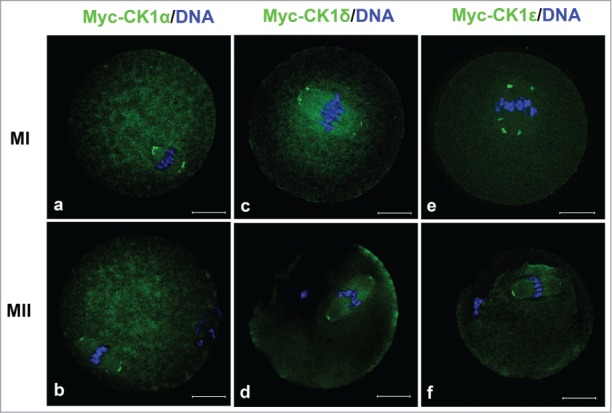
Figure 2.

The localization of myc-CK1α (a, b), myc-CK1δ (c, d) and myc-CK1ϵ (e, f) at MI and MII stages. GV oocytes were microinjected with myc-CK1α, myc-CK1δ or myc-CK1ϵ mRNA respectively and cultured to MI and MII stage, then fixed and stained with anti-myc antibody (green) and Hoechst 33342 (blue). Bar=20 μm.
