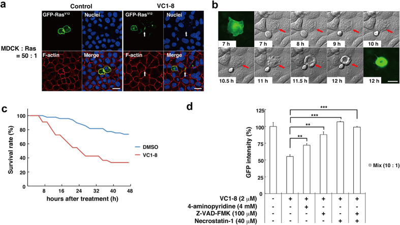
Figure 3. VC1-8 induces cell death of transformed cells that are surrounded by normal cells.
(a) Immunofluorescence images of MDCK-pTR GFP-RasV12 cells that are surrounded by MDCK cells in the absence (left) or presence (right) of 2 μM VC1-8 for 16 h. Cells are stained with Hoechst 33342 (blue) and Alexa-Fluor-568-conjuated phalloidin (red). Arrows indicate a cell with fragmentation. Scale bars: 20 μm. (b) Images of a representative time-lapse analysis of MDCK-pTR GFP-RasV12 cells that are surrounded by MDCK cells (red) in the presence of 2 μM VC1-8. Scale bar: 20 μm. (c) A graph showing the survival rate of MDCK-pTR GFP-RasV12 cells surrounded by MDCK cells in the presence of DMSO (blue) or 2 μM VC1-8 (red). n = 49 cells (DMSO) and 33 cells (VC1-8). The effect of VC1-8 was statistically significant (P < 0.0001). (d) Effect of apoptosis inhibitor (4-aminopyridine, Z-VAD-FMK) and/or necroptosis inhibitor (Necrostatin-1) on MDCK-pTR GFP-RasV12 cells mixed with MDCK cells. Data are mean ± SD from three independent experiments. Values are expressed as a ratio relative to DMSO treatment. **P < 0.01, ***P < 0.001.

