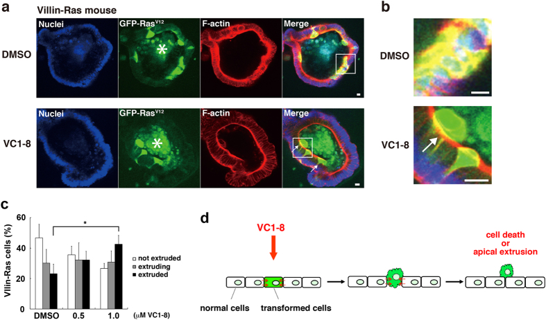
Figure 5. VC1-8 promotes the elimination of RasV12-transformed cells from the mouse intestinal epithelium.
(a) Immunofluorescence images of RasV12- transformed cells in the intestinal epithelium of the mouse crypt ex vivo culture in the absence (top) or presence (bottom) of 1 μM VC1-8. Intestinal epithelial cells were collected from mice obtained by crossing loxP-stop-loxP-KrasV12-IRES-eGFP and Villin-Cre/ERT2 mice. After applying them into the organ crypt culture, tamoxifen was added to the grown crypts to induce a mosaic expression of RasV12 within the mouse intestinal epithelium. Cells are stained with Hoechst 33342 (blue) and Alexa-Fluor-568-conjuated phalloidin (red). Arrows indicate the apically extruded GFP-RasV12-expressing cells from the epithelium. The asterisks indicate mucin-rich autofluorescent materials in the apical lumen. Scale bar: 50 μm. (b) A magnified image of the apically extruded RasV12-transformed cells (from a white box in (a)). (c) Effect of VC1-8 on the frequency of apical extrusion of RasV12-transformed cells in the ex vivo system: not extruded (white bar), extruding (grey bar) or extruded (black bar) RasV12-transformed cells from an epithelial monolayer; extruding: cells moving apically but still attached to the basal membrane, extruded: cells apically extruded and detached from the basal membrane. Data are mean ± SD from three independent experiments. *P < 0.05. (d) A schematic model of the cell competition-promoting effect of VC1-8. VC1-8 enhances the interaction between normal and transformed epithelial cells: promoting the attacking force of normal cells against transformed cells or attenuating the defensive force of transformed cells, eventually leading to cell death or apical extrusion of transformed cells.

