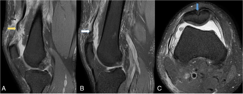
Figure 2.

(A) T2 proton density fat saturation MRI of the knee showing (A) medial sagittal image of complete rupture of vastus medialis and vastus intermedius tendon with proximal retraction (yellow arrow). (B) Lateral sagittal image showing intact lateral most fibres of vastus lateralis. (C) Axial image showing a groove over the patella indicating the patella bone plug harvest site (blue arrow).
