Fig. 2.

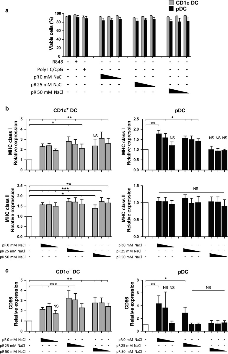
Protamine–RNA complexes are well tolerated by DCs and induce upregulation of maturation markers and MHC complexes. Purified CD1c+ DCs and pDCs were cultured 18–24 h with 15, 7.5, or 1.5 µg/ml of protamine–RNA complexes (pR) formed in 0, 25, or 50 mM NaCl. Untreated CD1c+ DCs or IL-3 treated pDCs were used as negative controls, while R848 was used as a control for TLR7/8 stimulation and poly I:C and CpG-C were used as positive controls for CD1c+ DCs or pDCs, respectively. a The cell viability was determined by flow cytometry. The mean percentage ± SEM of cells negative for live–dead marker from 7–8 CD1c+ DC donors and 6–8 pDC donors is depicted. b, c The relative expression of MHC class I and HLA-DR (b) and CD86 (c) on viable cells was calculated by normalizing the MFI values for each donor against the negative control. The fold increase ± SEM of 6–10 CD1c+ DC and pDC donors is depicted. Wilcoxon matched-pair signed-rank tests were performed on raw data, comparing against negative control, and are indicated by *(p < 0.05), **(p < 0.01), ***(p < 0.001), or NS (non-significant)
