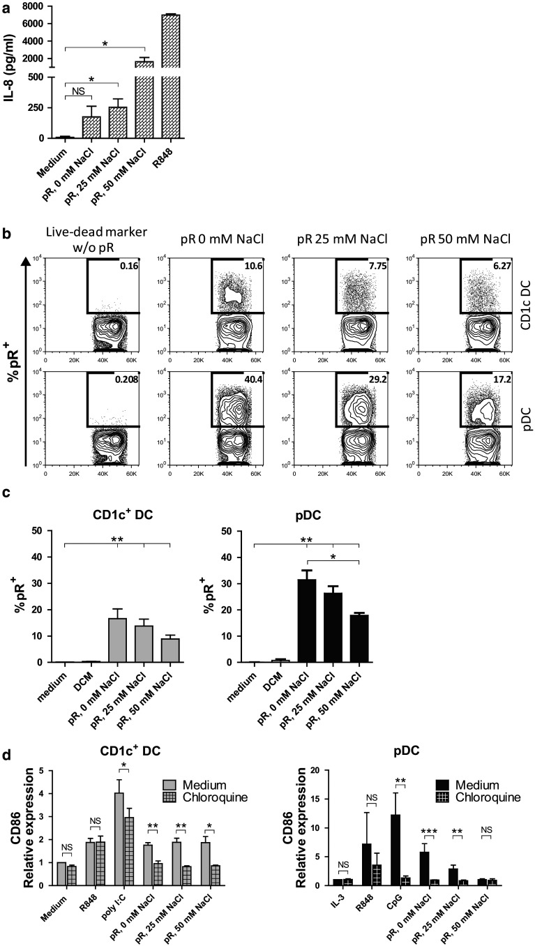
Fig. 4.
Effect of protamine–RNA complexes is TLR mediated. a TLR8 expressing HEK cells were cultured overnight with medium alone, R848, or protamine–RNA complexes (pR) formed in 0, 25, or 50 mM NaCl. The mean results ± SEM from three individual experiments run in triplicate or duplicate is depicted. T tests were performed on pooled data between indicated groups and are indicated by *(p < 0.05) or NS (non-significant). (b-c) DCs were cultured for 1 h with live–dead marker-labeled protamine–RNA complexes (pR) formed in 0, 25, or 50 mM NaCl or with live–dead marker alone and analyzed by flow cytometry. Percentages of protamine–RNA-positive cells were calculated on gated DCs and depicted as a representative figure (b) or as the mean uptake ± SEM from 7 CD1c+ DC and pDC donors (c). Wilcoxon matched-pair signed-rank tests were performed between indicated groups and are indicated by *(p < 0.05) or **(p < 0.01). d CD1c+ DCs and pDCs were pre-incubated for 1 h with chloroquine before the addition of medium alone or IL-3, R848, poly I:C or CpG-C, or protamine–RNA complexes (pR) formed in 0, 25, or 50 mM NaCl. The upregulation of CD86 was measured by flow cytometry after overnight culture and the relative expression was calculated by normalizing the MFI values for each donor against the negative control. Fold increase ± SEM of 4–5 CD1c+ DC and 3–5 pDC donors is depicted. T tests were performed on raw data between indicated groups and are indicated by *(p < 0.05), **(p < 0.01), ***(p < 0.001), or NS (non-significant)

