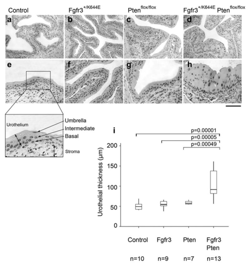
Figure 1. Increased thickness of the UroIICreFgfr3+/K644EPtenflox/flox urothelium.
Representative images of H&E stained bladder sections of Control (a, e), UroIICreFgfr3+/K644E (b, f), UroIICrePtenflox/flox (c, g) and UroIICreFgfr3+/K644E Ptenflox/flox (d, h) at low (a-d) and high magnification (e-h). The murine urothelium consists of three layers, namely umbrella, intermediate, and basal cells and borders with connective tissue and the stroma (e, insert). Scale bar represents 200 μm in panel a-d and 100 μm in panel e-h. Thickness of the urothelium was quantified in Control, UroIICreFgfr3+/K644E (Fgfr3), UroIICrePtenflox/flox (Pten) and UroIICreFgfr3+/K644EPtenflox/flox (Fgfr3Pten) in the number of animals indicated (i). The error bars indicate the standard deviations.

