
Figure 2.
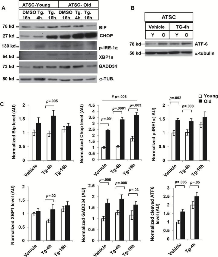
Effect of thapsigargin (Tg) on adipose tissue derived stromal cells from young and old mice. (A) Western blot analysis of endoplasmic reticulum (ER) stress response pathway components: Adipose tissue stromal cells (ATSCs) from young (5 mice) and old mice (3 mice) were treated with Tg (330nM) and the lysates were subjected to Western blot analysis with BIP, CHOP, p-IRE1α, XBP-1, GADD34, and ATF-6 antibodies (B). Elevated expression of these ER stress responses was observed in old ATSC both in resting and Tg-induced condition. (C) The relative density of protein bands for BIP, CHOP, p-IRE-1a, XBP-1, GADD34, and ATF-6 were plotted after normalization with γ-tubulin intensity. Values were presented as mean ± SD and p < .05 in Student’s t test was considered significant. # p value indicates the significance level of two-way analysis of variance for the interaction between treatment (Tg) and age factor (y vs o).