Fig. 3.

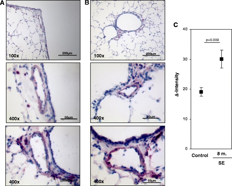
S100A4 protein expression in intrapulmonary vessels in mouse lungs after smoke-exposure. Representative images of intrapulmonary arteries from a control animals, b smoke exposed animals. Bars represent 50 μm and 200 μm. c semi-quantitative analysis of S100A4 protein expression in intrapulmonary arteries. Δ Intensity- color intensity of S100A4 protein after immunohistochemical staining. 8 m-eight months, SE-smoke exposure
