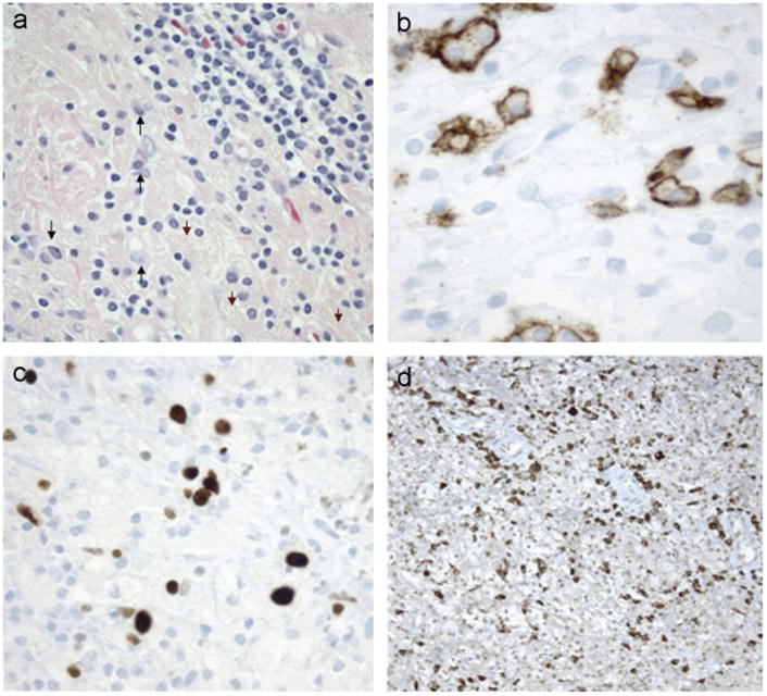
Fig. 3.

Biopsy Confirming Malignant Transformation. (a)–(d) Brain biopsy 5/07. A third brain biopsy demonstrates an increase in atypical plasma cells (arrows) with crystal laden macrophages (arrowheads) and lymphocytes (a). CD138 is positive in plasma cells with enlarged, atypical nuclei (b). Ki67 demonstrates proliferation of atypical lymphoplasmacytes with enlarged nuclei (c). Immunocytochemistry for κ-light chains highlights many lymphocytes/plasma cells (d). Findings are consistent with plasma cell neoplasm associated with CSH.
