Fig. 3.
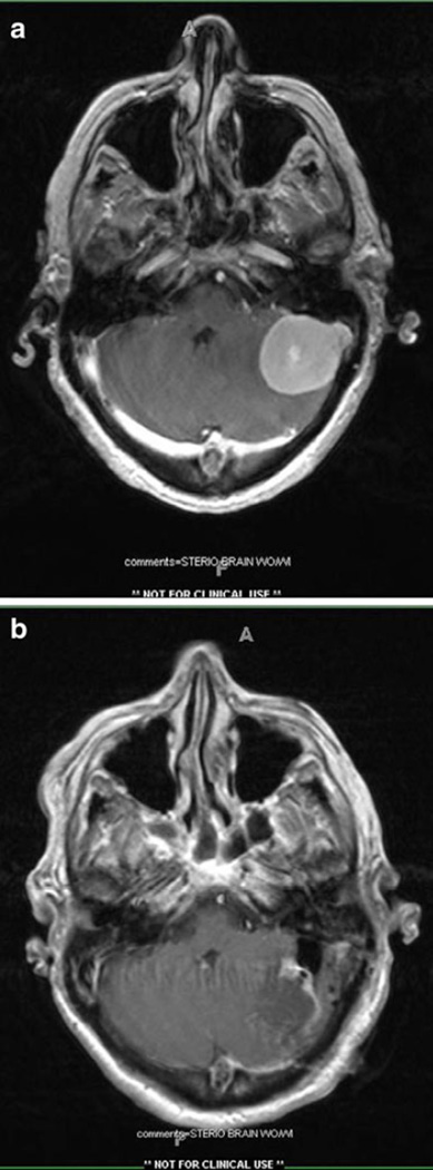
a Pre-operative magnetic resonance imaging (T1-weighted with contrast) for case illustration (see text). Noted is an extra-axial homogeneously enhancing mass along the transverse–sigmoid sinus junction. b Post-operative MR scan from case illustration demonstration complete resection of previously described mass

