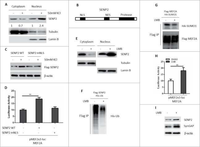
Figure 2.
Activity-dependent stimuli promote SENP2 nuclear translocation. (A) SHSY5Y cells were treated with 50 mM KCl or 150 mM NaCl for 2 h and the nuclear-cytoplasmic fraction of these cells was conducted. Lysates from each fraction were immunoblotted for SENP2. Tubulin and Lamin B were used to indicate cytoplasmic or nuclear fraction, respectively. The number on the bottom of Western blot lanes indicated signal intensity of SENP2 protein against β-Tubulin or Lamin B. The signal intensity of SENP2 protein against β-Tubulin or Lamin B without KCl treatment was set as 1. (B) Schematic diagrams of the NLS, NES and Protease domain of SENP2. (C) SHSY5Y cells were transfected with Flag-SENP2 WT or Flag-SENP2 mNLS for 36 h. Cells were treated with 50 mM KCl or 150 mM NaCl for 2 h. The whole cell lysates were immunoblotted with Flag antibody. (D) SHSY5Y cells were transfected with indicated plasmids for 36 h. Cells were then treated with 50 mM KCl or 150 mM NaCl for 2 h, the luciferase activity was measured. The y axis represents normalized luciferase activity ± SEM (n = 3). **represents P < 0.01. (E) SHSY5Y cells were transfected with Flag-SENP2 for 32 h and treated with 10 nM LMB or DMSO for 2 h and the nuclear-cytoplasmic fraction of these cells was conducted. Lysates from each fraction were immunoblotted for Flag-SENP2. Tubulin and Lamin B were used to indicate cytoplasmic or nuclear fraction, respectively. (F) SHSY5Y cells were transfected with Flag-SENP2 and His-ubiquitin plasmids for 36 h. Cells were then treated with 10 nM LMB. Cells were lysed and subjected to immunoprecipitation with Flag M2 beads. Immunoprecipitated proteins were immunoblotted with anti-His antibody. (G) SHSY5Y cells were transfected with Flag-MEF2A and HA-SUMO1 for 36 h. Cells were then treated with 10 nM LMB for 2 h. Cells were lysed and subjected to immunoprecipitation with Flag M2 beads. Immunoprecipitated proteins were immunoblotted with anti-HA and Flag antibodies. (H) SHSY5Y cells were transfected with MEF2A and MEF-luciferase reporter plasmids for 36 h. Cells were then treated with 10 nM LMB for 2 h, the luciferase activity was measured. The y axis represents normalized luciferase activity ± SEM (n = 3). **represents P < 0.01. (I) Lysates of primary rat cortex neuron cells treated with 10 nM LMB or DMSO were immunoblotted for SynGAP and SENP2.

