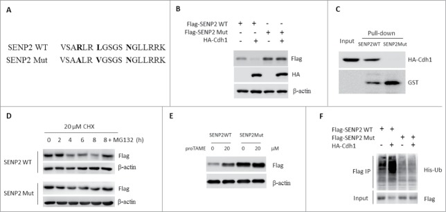
Figure 5.

D-box-dependent degradation of SENP2 by APCCdh1. (A) Schematic diagrams of SENP2 WT and Mut. (B) SHSY5Y cells were transfected with indicated plasmids for 48 h. The whole cell lysates were immunoblotted with Flag and HA antibodies. (C) Beads coated with bacterially expressed GST-SENPWT or GST-SENP2Mut were incubated with purified HA-Cdh1 protein. Beads were washed, and the bound proteins were analyzed by western blotting with indicated antibodies. (D) SHSY5Y cells transfected with Flag-SENP2 WT or Flag-SENP2 Mut for 36 h were treated with 20 μM CHX for the indicated time. In MG132 group, 10 μM was added 6 h before cell harvested. The whole cell lysates were immunoblotted with indicated antibodies. (E) SHSY5Y cells transfected with Flag-SENP2 WT or Flag-SENP2 Mut for 36 h were treated with 10 μM TAME for 12 h. The whole cell lysates were immunoblotted with indicated antibodies. (F) 293T cells were transfected with indicated plasmids for 36 h. Cells were lysed and subjected to immunoprecipitation with Flag M2 beads. Immunoprecipitated proteins were immunoblotted with indicated antibodies.
