
Figure 1.
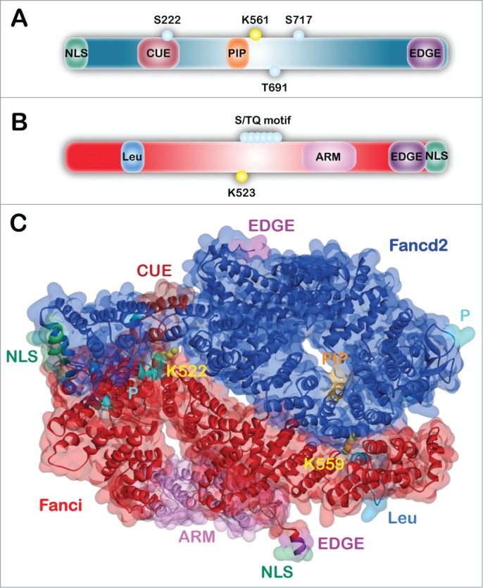
Domain architecture and structure of FANCD2 and FANCI. (A) Schematic of the FANCD2 protein indicating the amino-terminal NLS (nuclear localization signal) domain (green), CUE (coupling of ubiquitin conjugation to endoplasmic reticulum degradation) domain (maroon), PIP-box (PCNA-interacting protein motif) (orange), and the C-terminal EDGE motif (purple). Functionally-characterized phosphorylation sites (teal) and K561 monoubiquitination site (yellow) are indicated by small circles. (B) Schematic of the FANCI protein indicating the Leu (leucine zipper) domain (light blue), ARM (armadillo repeat) domain (pink), and C-terminal EDGE motif (purple) and NLS domain (green). The S/TQ motif (teal) and K523 site of monoubiquitination (yellow) are indicated by small circles. (C) Mouse Fanci-Fancd2 heterodimer crystal structure represented as both surface and ribbons with domains indicated. This structure was solved by the Pavletich group in 2011(PDB ID: 3S4W).35