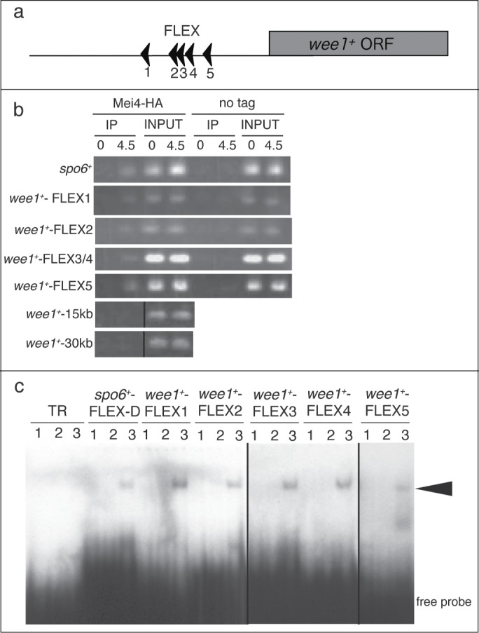
Figure 2.

Mei4p binds to the FLEX sequences upstream of wee1+ both in vivo and in vitro. (A) Schematic representation of the positions of the FLEX sequences associated with wee1+. The arrowheads indicate the direction of the FLEX sequence from 5′ to 3′. ORF, open reading frame; (B) A chromatin immunoprecipitation assay of FLEX sites associated with wee1+ was performed with antibodies against HA or control IgG at the indicated times after induction of meiosis in cells expressing (Mei4-HA) or not expressing (no tag) mei4+-HA. wee1+-15kb and wee1+-30kb were about 15 kb and 30 kb downstream, respectively, of the start codon of wee1+ and were used as negative controls. TheFLEX-D site of spo6 was used as a positive control. The images of the immunoprecipitates of wee1+-15kb and wee1+-30kb are different from those of the inputs, but the experimental conditions were the same; (C) Electrophoretic mobility shift assay analysis of wee1+ FLEX sites with a recombinant glutathione S-transferase (GST)-Mei4p fusion protein. Buffer only (lane 1), GST (lane 2), or purified recombinant GST-Mei4p (71–182) (lane 3) were incubated with the indicated labeled oligonucleotide probe. The TR probe is unrelated to the FLEX sequence and was used as a negative control. The arrowhead indicates shifted bands. The image of wee1+−FLEX3 and wee1+−FLEX4 is different from the rest of the image, but the experimental conditions were the same.
