Figure 2.

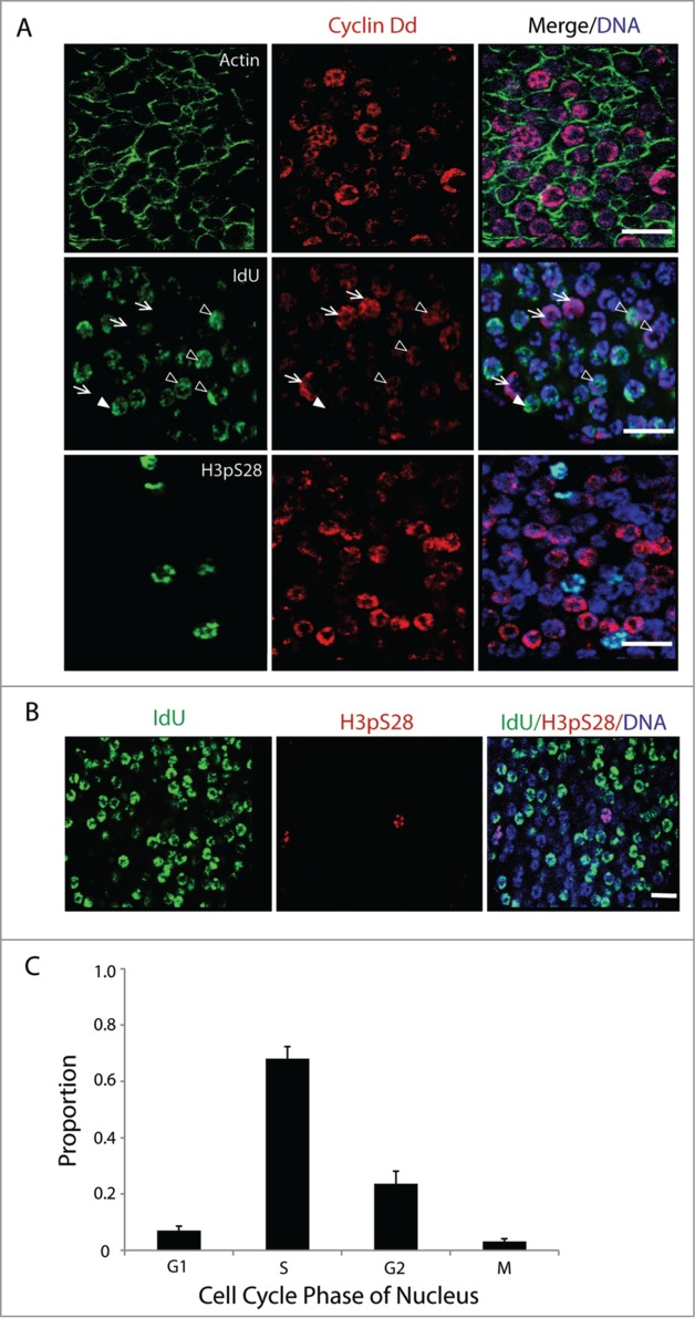
Asynchronous proliferation of germ nuclei with a distinct G1-phase. (A) Immunostaining of asynchronous mitotic germ nuclei show different levels of Cyclin Dd expression (n = 10 animals). Actin network within the single-cell coenocyst is stained in green. Cyclin Dd expression peaks in G1 (arrows), persisted at reduced levels in early S (arrowheads; Campsteijn et al.24) and was absent thereafter. Cyclin Dd expression was not seen in nuclei expressing the mitotic marker H3pS28. (B) Larger overview of germline nuclei indicating proportions in G- (no staining), S- (IdU incorporation) and M-phase (H3pS28 staining) prior to meiotic entry. (C) Proportion of germ nuclei in each of the proliferative cell cycle phases was assessed (n = 10 animals) by immunostaining in combination for markers: Cyclin Dd (G1), short IdU pulse (S), and H3pS28 (M). Nuclei that were negative for all of these markers were in G2. Error bar indicates standard error. Scale bars = 10 µm.
