Figure 3.

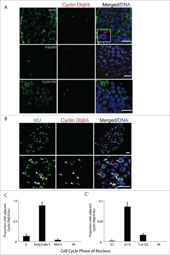
Cyclin Dbβ/δ cytoplasmic foci and the G1/S transition in germline nuclei. (A) Immunostaining of mitotic germline nuclei with antibodies specific to C-terminal regions of Cyclin Db splice variants β/δ. Cyclin Dbβ/δ were present as foci in the syncytial germline cytoplasm adjacent to nuclei. These foci were not observed adjacent to nuclei in M-phase (H3pS28 staining) and were also not preferentially associated with nuclei staining most intensely for Cyclin Dd (n = 10 animals). Inset depicts magnification of a cytoplasmic Cyclin Dbβ/δ focus adjacent to a nucleus. (B) Cytoplasmic foci of Cyclin Dbβ/δ were adjacent to nuclei with low amounts of IdU incorporation (arrows) but were not adjacent to nuclei with no IdU incorporation (G-phase; empty arrowheads) or nuclei with large amounts of IdU incorporation (filled arrowheads). (C) Cyclin Dbβ/δ foci were selected (n = 10 animals; 1000 foci) and the cell cycle phase of the closest nucleus was assessed based on extent of IdU incorporation and H3pS28 staining. (C') Reciprocally, nuclei in G1 (no incorporation of IdU, Cyclin Dd staining), G1/S (weak incorporation of IdU, weak Cyclin Dd staining) S or G2 (increased IdU staining, no Cyclin Dd staining) or M (H3pS28 staining) were selected (n = 10 animals; 900 nuclei) and assessed for the presence or absence of adjacent Cyclin Dbβ/δ foci. Error bars indicate standard errors. Scale bars = 10 µm.
