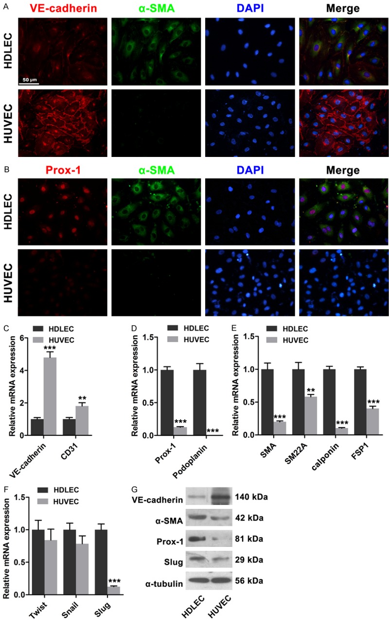
Figure 1.

Mesenchymal status of HDLECs compared to HUVECs. A. Immunofluorescence showed lower expression of VE-cadherin and higher expression of α-SMA in HDLECs compared to HUVECs. B. Immunofluorescence showed higher expression of Prox1 in HDLECs. C. Endothelial markers VE-cadherin and CD31 were lower in HDLECs. D. Lymphatic endothelial markers Prox1 and Podoplanin were higher in HDLECs. E. Mesenchymal proteins including α-SMA, SM22α, SM and FSP-1 were higher in HDLECs. F. Real time PCR assays revealed the expression of Twist, Snail and Slug in HDLECs and HUVECs. G. Western blots showed the expression levels of VE-cadherin, α-SMA, Prox-1 and Slug in HDLECs compared to HUVECs. α-Tubulin was used as loading control. All data are presented as mean ± SE from three different experiments with duplicate. *, P < 0.05; **, P < 0.01 versus HDLECs.
