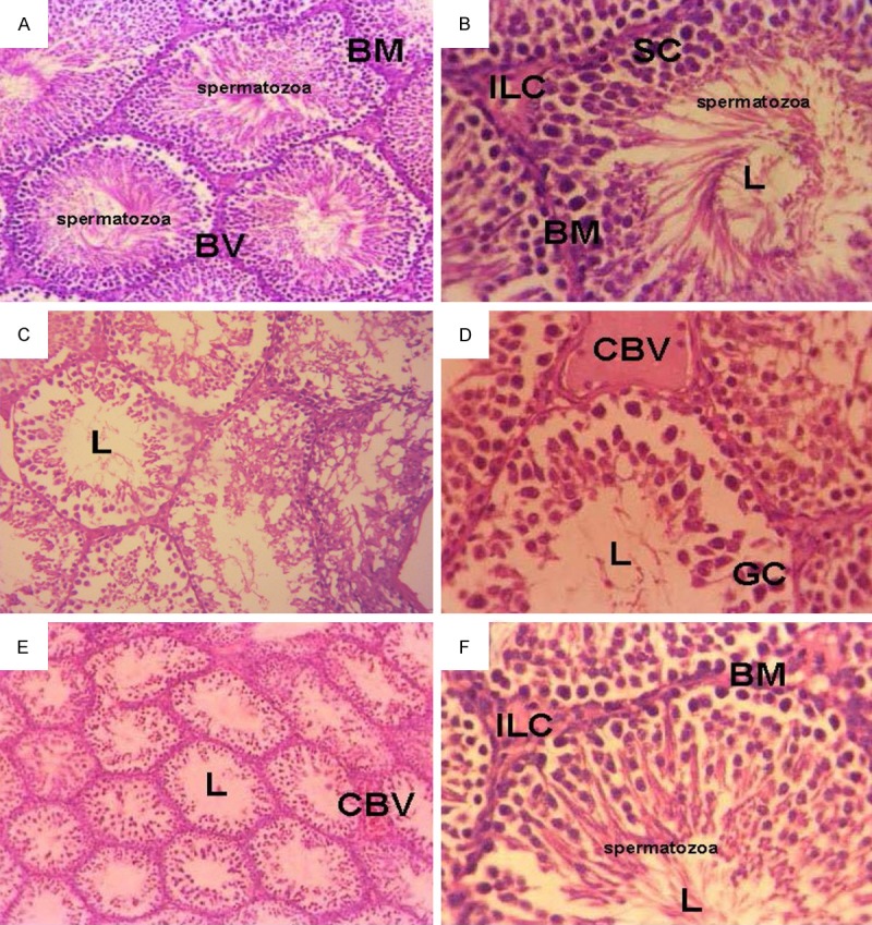
Figure 4.

Micrograph of transverse section of testis of (A) control rats showed normal seminiferous tubule. The seminiferous tubule has well-developed basement membrane (BM), blood vessel (BV), numerous sertoli cells, spermatogonium, and spermatocytes in the lumen (L) of the tubule. (H&E × 200). (B) Rats injected with MOE, showed normal arrangement of seminiferous tubules which have numerous sertoli cells (SC), interstial Leydig cells (ILC) and different stages of the spermatocytes (H&E × 400). (C-E) Testes of rats treated with EMR, showed destructed seminiferous tubules associated with degenerated spermatogonic cells, giant cells (GC), congested blood vessel (CBV), absent interstitial leyding cells and free tubule lumen (L) spermatocytes (H&E × 400). (F) Testis of rats treated with EMR and MOE for 8 weeks showed the normal arrangement of seminiferous tubule cells which have numerous cells sertoli spermatogonium, spermatocytes (H&E × 400).
