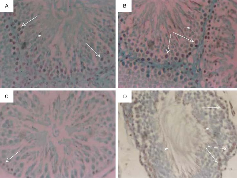
Figure 5.

Micrograph of immunohistochemical analysis of paraffin-embedded testis using PCNA showing brown nuclear staining (A) control, (B) MOE injected animal group, (C) EMR exposure animal group and (D) EMR exposure animal treated with MOE group. Positive staining in spermatogonia (white arrows) and early-stage spermatocytes (*). (immunoperoxidase × 250).
