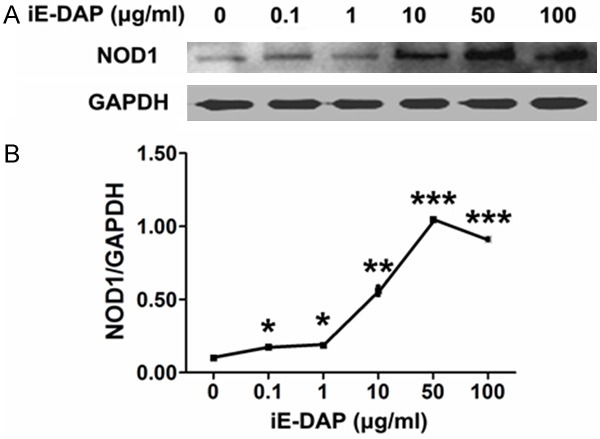
Figure 2.

iE-DAP triggered NOD1 expression of leuk-1 cells in a dose-dependent manner. A. Representative immunoblot bands of NOD1. B. Density analyses of bands indicated that treatment of cells with 0.1, 1, 10, 50 and 100 μg/ml iE-DAP for 24 h clearly enhanced NOD1 expression. Following 50 μg/ml iE-DAP treatment for 24 h, NOD1 expression reached the peak level. Density data of bands were represented as means ± SE (n = 3). Statistical significance: *P < 0.05, **P < 0.01, ***P < 0.001, vs. control.
