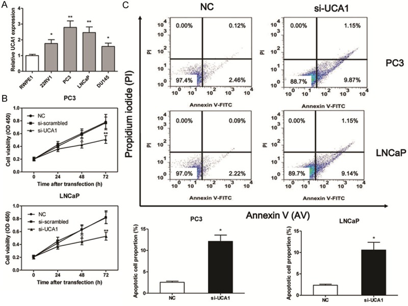
Figure 3.

UCA1 loss-of-function induces cell apoptosis. The expression of UCA1 in prostate cancer cell lines (A). The cell viability is measured by CCK8 in PC3 and LNCaP cell lines after si-UCA1 transfection (B). The apoptotic cell proportion is measured by flow cytometric analysis of annexin V/PI double staining after the si-UCA1 transfection for 48 h, upper right and lower right in the cell distribution pictures represent the late and early apoptotic cells proportion respectively (C). Values are expressed as mean ± SD, *P < 0.05, **P < 0.01 versus control group. N = 3 in each group.
