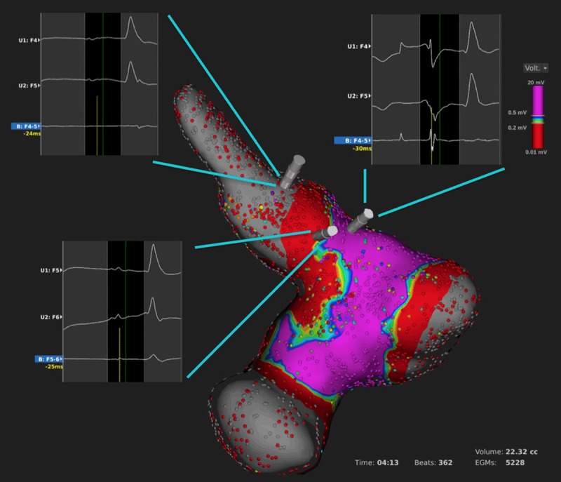
Figure 5.

LA and PV voltage map after complete isolation of the upper PV and verification of the ablation lesions in the review mode. Please note the different unipolar and bipolar electrograms within the PV (left upper inlet), along the ablation line (left lower inlet) and outside the ablation line (right upper inlet).
