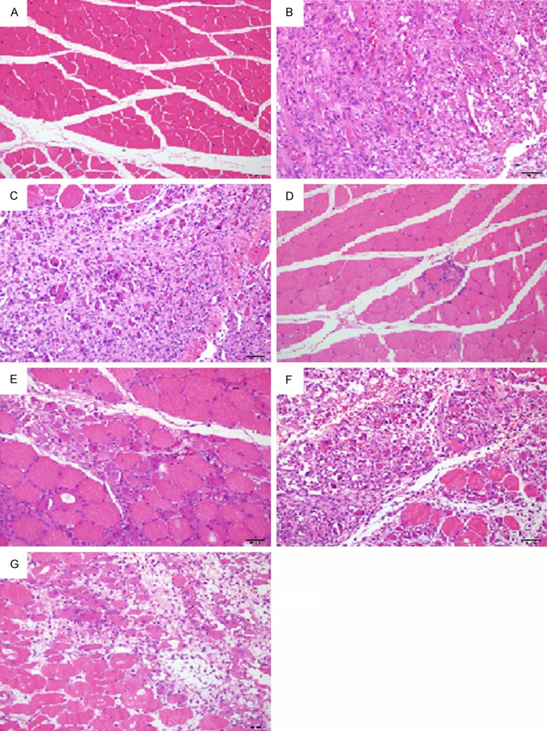
Figure 4.

Representative histopathological micrographs (× 200, HE staining) in the injured muscle of rats with the control (A), model (B), vehicle treatment (C), high or low-dose Xiangqing anodyne spray treatment (D, E), Cynanchum paniculatum spray (F) or Illicium henryi spray (G) treatment. Normal myocyte in (A); Severe myocyte degeneration and necrosis following marked fibrosis, massive inflammatory cell infiltrates and interstitial ecchymosed in (B, C); A little area of the degeneration and necrosis in (D); Markedly reduced area of the degeneration and necrosis in (E); Markedly difference in (F, G) compared to (B) was normal myocyte shown in the necrosis area.
