
Figure 2.
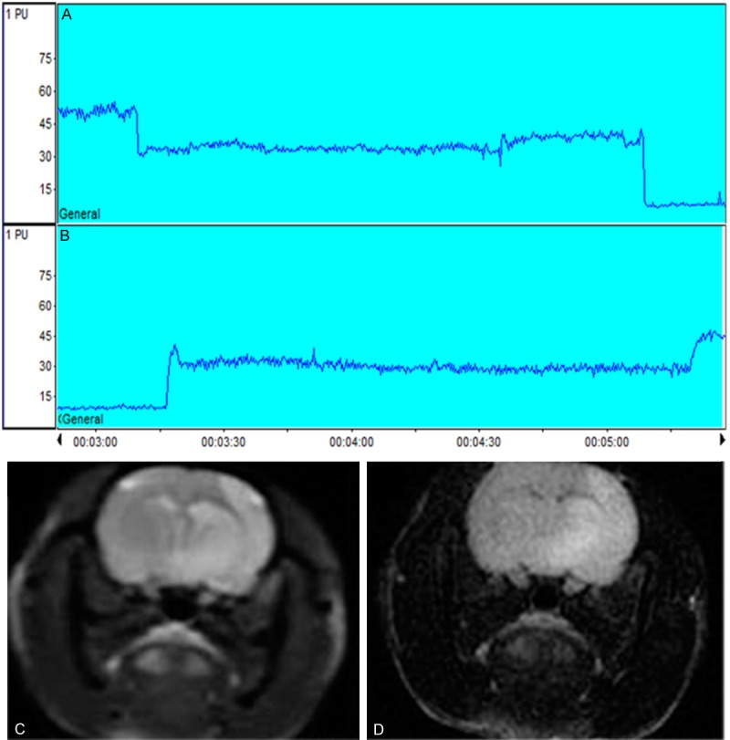
Representative cerebral blood flow and magnetic resonance imaging of MCAO/R model of rat. A, B. Laser Doppler Flowmetry showed that the CBF was two drops from baseline during occlusion procedure and two jumps during reperfusion procedure, the percentage drop from baseline ≥80%. C, D. Brain edema accompanying focal ischemia is visualized in T2-weighted and T2-Flair MRI images.