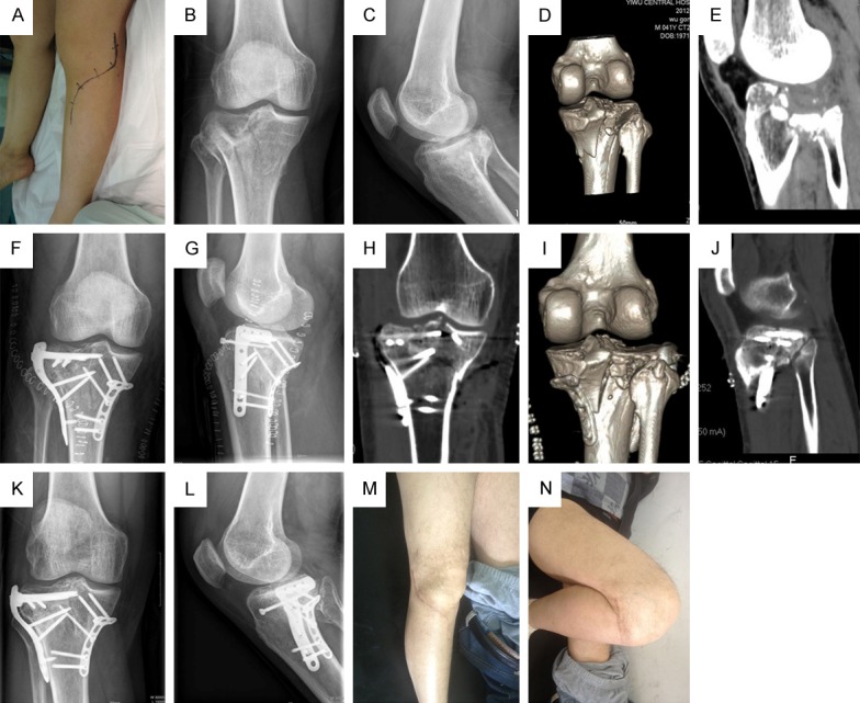
Figure 1.

A: Incision of the extended anterolateral approach; B, C: Anteroposterior and lateral x-ray image of posterolateral tibial plateau fracture before surgery; D, E: CT films of obviously posterolateral tibial plateau fracture before surgery; F, G: Anteroposterior and lateral x-ray image of satisfied reduction of posterolateral tibial plateau fracture via extended anterolateral approach after surgery; H-J: CT films of anatomical reduction of posterolateral tibial plateau fracture after surgery; K, L: No fracture displacements during one year follow-up period after surgery; M, N: Satisfied flex function in the re-examination one year later after surgery.
