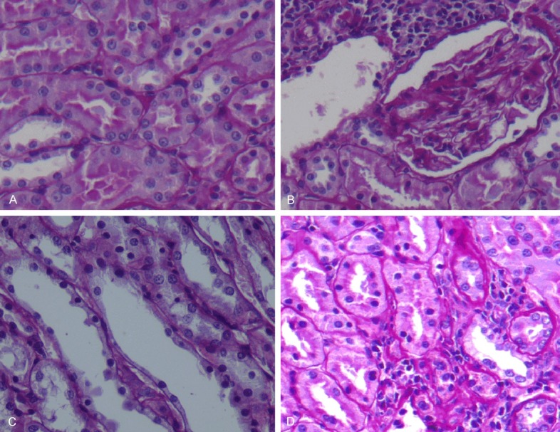
Figure 1.

(A) Control group, (B, C) Mtx group, (D) Mtx-O3 group. (A) Control group shows normal kidney architecture. (B) In Mtx group interstitial mononuclear inflammation was seen also note that glomerules are shrunken and tubules are athropic. Glomerular and tubular basal membrane wrincling is seen. (X20, PAS). (C) Tubular cell loss; several tubular cell nuclei are not seen along this tubule. Also not picnotic nucleus reflects apoptotic cell necrosis. (X40, PAS). (D) Moderate degeneration on glomerulus and tubules was seen.
