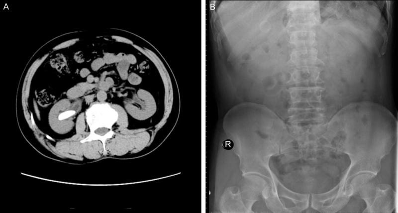
Figure 3.

Male patient, 69 year-old. A: CT scanning showed an irregular mass measuring 32 mm × 13 mm in size with higher density. B: The shape of both kidneys did not show up clearly in the KUB radiography; a radiolucent shadow with the size of 30 mm × 13 mm was observed in the right kidney; no evidence of radiopaque calculus shown in the left kidney.
