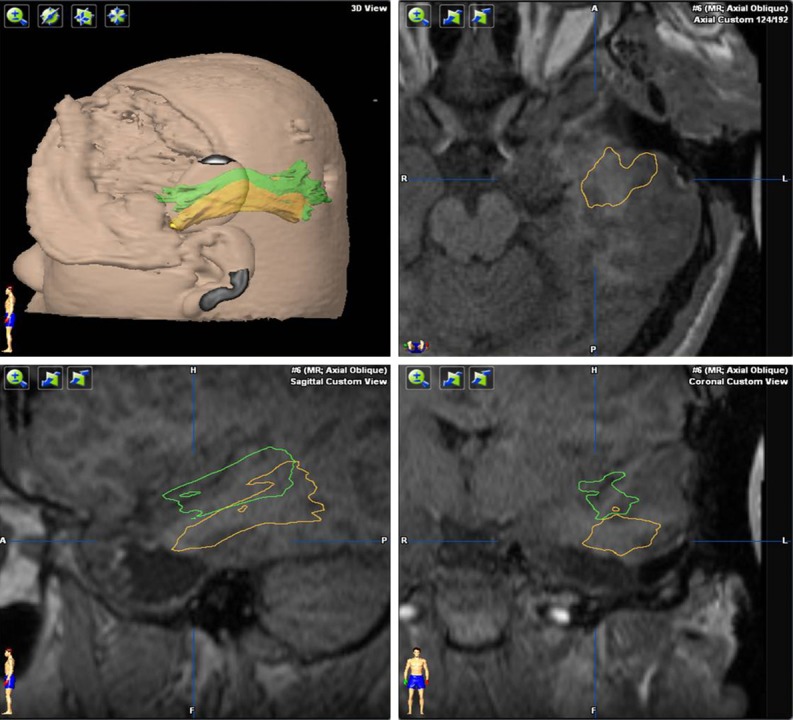
Figure 3.

Fusion of preoperative and intraoperative magnetic resonance imaging (MRI) scans. After removing the anterior temporal lobe according to the projection of the optic radiation onto the cortex, an intraoperative MRI scan was performed and the optic radiation was reconstructed (green). We then judged whether the residual temporal lobe tissue needed to be removed. The intraoperative MRI scan was then fused to the preoperative MRI. Comparing the intraoperative optic radiation (green) with the preoperative optic radiation (yellow), we found that shifting of the brain because of cerebrospinal fluid movement during surgery was very common. The temporal lobe usually shifted toward the center line and the top of the head, but there was very little shift from front to back if the patient’s head was positioned on the side.
