
Figure 5.
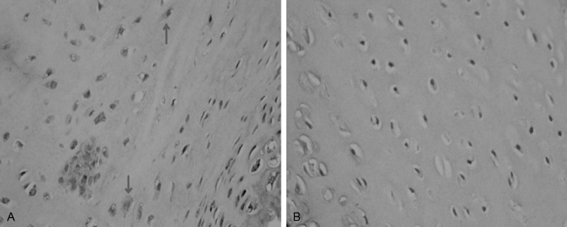
A: Uneven expression of type II collagen in the ECM in experimental group, cells were surrounded with hyperchromatic, wavy or irregular-shaped collagen in ECM, and obvious hyperchromatic cytoplasm showed in chondrocyte (indicated with arrow); Magnification × 400. B: lower staining of ECM of normal chondrocytes in control group, lighter staining in cytoplasm and fewer stained cells. Magnification × 400.