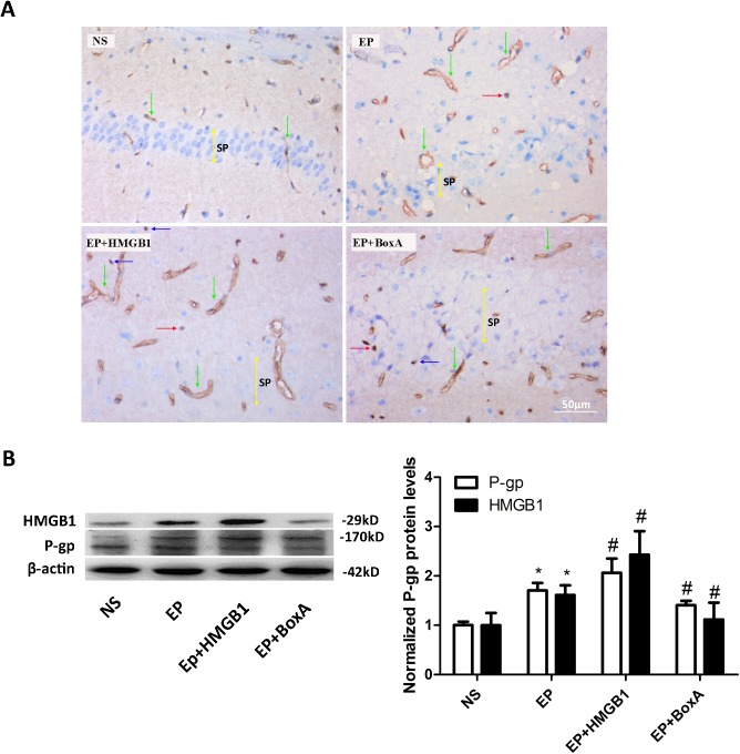
Fig 2. Pre-treatment with HMGB1 enhanced KA-induced up-regulation of P-gp in the epileptic brain.
(A) Immunohistochemical staining of P-gp in CA1 hippocampal areas of each group mice (NS group, n = 5; EP group, n = 6; EP+BoxA group, n = 6; EP+BoxA group, n = 6). HMGB1 injection enhanced the over-expression of P-gp in the EP group mice while BoxA attenuated it. Scale bar: 50 μm. NS: normal saline control group; EP: KA-induced epileptic seizure group; EP+HMGB1: EP group pretreated with HMGB1; EP+BoxA: EP group pretreated with BoxA. Green arrows: vascular endothelial cells; Red arrows: neurons; Blue arrows: glial cells; Yellow bidirectional arrows: Stratum pyramidale (sp). (B) Western blotting detects the protein levels of HMGB1 and P-gp in the mice brains of each group 24 h after seizure onset (B left panel, n = 6). Protein levels were quantified and normalized to that in the NS group (B right panel). Injection of HMGB1 and BoxA increased and decreased the protein levels of HMGB1 and P-gp in the EP group mice respectively. *P<0.05 vs. NS group; # P<0.05 vs. EP group. HMGB1, high-mobility group box-1; KA, kainic acid; P-gp, P-glycoprotein.

