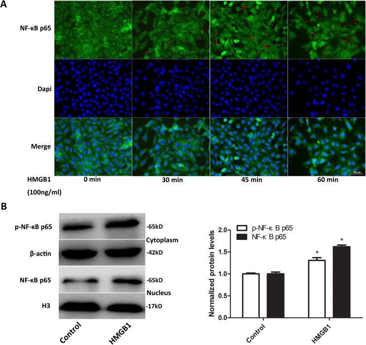
Fig 5. HMGB1 enhanced phosphorylation and nuclear translocation of NF-κB p65 in bEnd.3 cells.
(A) NF-κB p65 subcellular distribution in bEnd.3 cells treated with HMGB1 for indicated durations was observed using immunofluorescence staining. HMGB1 promoted cytoplasmic to nuclear translocation of NF-κB p65 in bEnd.3 cells. Representative pictures are shown. Scale bar: 100 μm. Red arrows: cells with nuclear NF-κB p65 staining. (B) Protein levels of phosphor-NF-κB p65 and NF-κB p65 in the cytoplasm and nuclear fractions from bEnd.3 cells treated with HMGB1 for 60 min. H3 and β-actin were utilized as loading controls for nuclear and cytoplasmic proteins, respectively. Protein levels were normalized to the cells treated without HMGB1. HMGB1 increased cytoplasmic p-NF-κB p65 and nuclear NF-κB p65 levels in bEnd.3 cells. Data were shown as mean±SD; n = 3. *P<0.05 vs. cells treated with medium only. HMGB1, high-mobility group box-1; NF-κB, nuclear factor-kappa B; SD, standard deviation.

