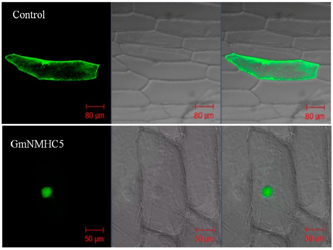
Figure 3.
Cellular localization of eGFP and GmNMHC5-GFP fusion proteins. Photographs were taken in a dark field for green fluorescence (left column) and a bright field for cell morphology (middle column). The right column is the overlapping view of the dark and bright fields for comparative clarity.

