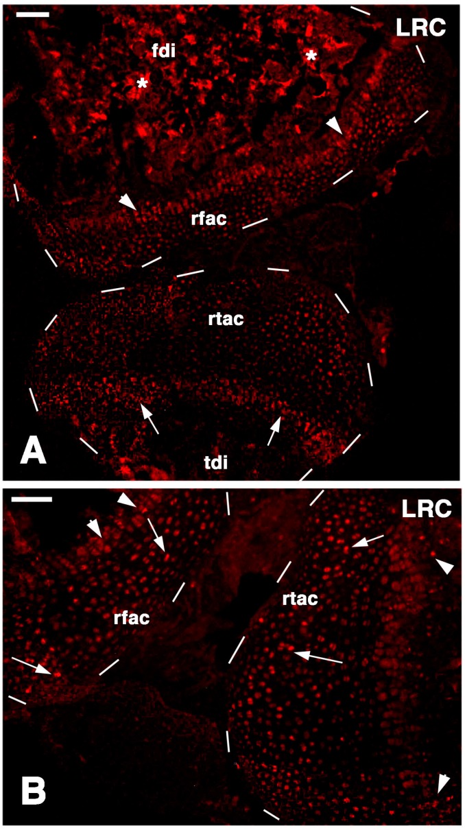
Figure 5.
TRITC immunofluorescence for LRCs in regenerated cartilaginous epiphyses at four-and-a-half weeks post-injury. (A) General view of the regenerated cartilaginous articular cartilage of the femur (rfac) and tibia (rtac), both outlined by dashes. Sparse labeled cells are present within the cartilage and in the metaphyseal growth plates (arrowheads in femur, arrows in tibia). Asteriks indicate a non-specific labeling of the mineralized matrix in the femur diaphysis (fdi) and tibia diaphysis (tdi). Bar, 50 µm; (B) detail of the regenerated femur articular cartilage (rfac) and that of the tibia (rtac), for which surfaces are outlined by dashes. Sparse intensely labeled LRCs are seen within the cartilaginous tissue (arrows) and few also in the metaphyseal growth plate (arrowheads). Bar, 50 µm.

