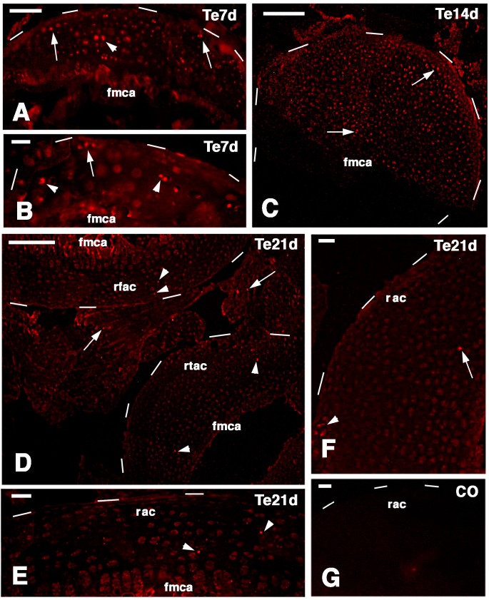
Figure 8.
TRITC immunolabeling for a telomerase-1 components (Te) in the regenerating epiphyses (dashes outline their surfaces). (A) At seven days few labeled cells are seen within the new cartilage (arrowhead) and along the surface (arrows) of a tibia. Bar, 50 µm; (B) other mass of cartilaginous tissue in a tibia at seven days post-injury showing labeled cells on the surface (arrow) and inside the cartilaginous tissue (arrowheads). Bar, 25 µm; (C) sparse intensely labeled cells within (arrowhead) and near the surface (arrow) of a cartilaginous epiphyses 14 days after injury. Bar, 100 µm; (D) Very sparse labeled cells are seen within the femur (top arrowheads) and tibia (bottom arrowheads) articular cartilages 21 days after injury. Few labeled cells (arrows) are also seen in the meniscus. Bar, 100 µm; (E) detail showing two labeled cells (nuclei, arrowheads) within the regenerated cartilage at 21 days. Bar, 25 µm; (F) other detail on labeled cells (arrow inside the cartilage; arrowhead near the surface) at 21 days post-injury. Bar, 25 µm; (G) control section. Bar, 25 µm. Legends: fmca, forming metaphyseal cartilage (plate); rfac, regenerated femur articular cartilage; rac, regenerated articular cartilage; rtac, regenerated tibia articular cartilage.

