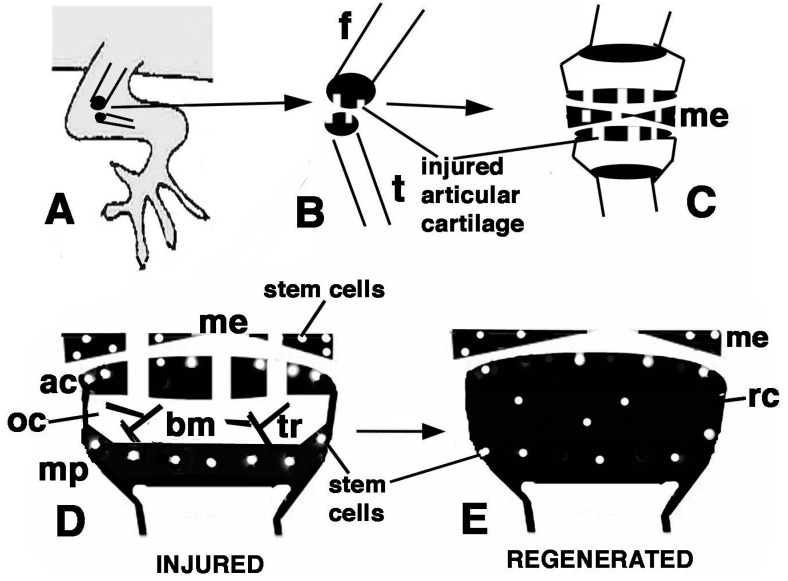
Figure 9.
Schematic drawing resuming the present observations. (A) depicts a lizard knee where the injury of the articular surfaces is indicated in (B); (C) depicts the damaged area (white) produced on the articular cartilages and meniscus of the knee; (D) further depicts the structure of an injured epiphyses and meniscus with the position of Long Retaining Cells (LRC)/telomerase positive and putative stem cells in the articular cartilage and metaphyseal plate indicated; (E) depicts that the regenerated cartilage (in black) occupies the entire epiphyses at 21−32 days (four-and-a-half weeks) post-injury while stem cells appear sparse and not localized in the surface or metaphyseal plate, and are also present in the meniscus. (ac, articular cartilage; bm, bone marrow; f, femur; me, meniscus; mp, metaphyseal (growth) plate; oc, ossification center; rc, regenerated cartilage; t, tibia; tr, bone trabeculae.)

