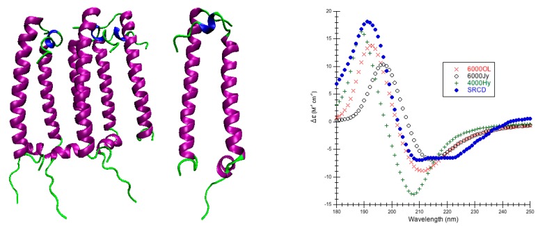
Figure 6.
Light-Harvesting Protein Complex II. (Left & Center) Secondary structure of light-harvesting protein complex II (PDB code 1NKZ [67]). The purple coils are helices (12–37, 40–46); the 310-helices are blue (6–8). The green coils are other structures. (Left) Asymmetric unit (A3B3); (Center) Heterodimer (AB). (Right) Predicted CD Using CDCALC. The 1NKZ AB dimer is minimized with 5000 conjugate gradient steps using NAMD/CHARMM22. Calculated spectra ignore all CH3 group hydrogens. The 6000 and 4000 refer to bandwidths in cm−1. Calculated spectrum show the smallest RMSD 6000 OL (), the largest RMSD 4000Jy (o), and an example helical parameter result, 4000 Hy (). The blue dots () are the experimental SRCD (CD0000114000) [44,59]. The CATH fold classification [53] is a combination of few secondary structures/irregular for chain A and mainly alpha/up-down bundle for chain B. Note: the complete hexameric asymmetric unit of the protein was not treated and neither were the any of the ligands (bacteriochlorophyll A, benzamidine, β-octylgucoside, rhodopin glucoside).

