Figure 3.
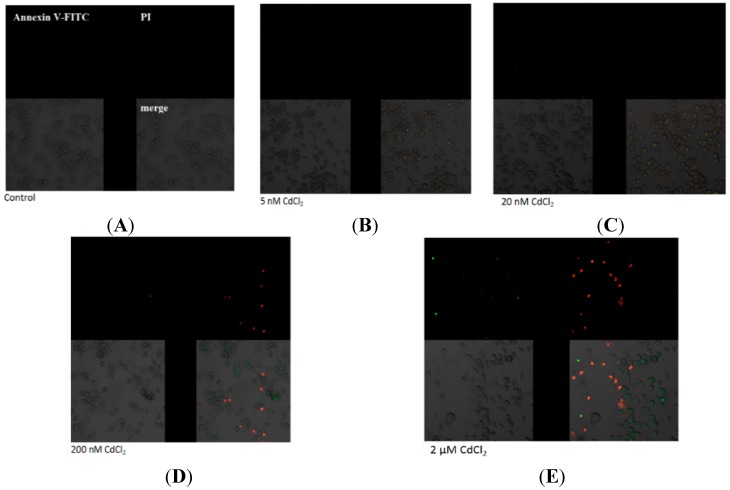
Imaging of apoptosis process by confocal microscopy in macrophages cultured with (A) control, cells incubated in RPMI 1640 medium with 10% FBS and H2O as vehicle; (B) 5 nM CdCl2; (C) 20 nM CdCl2; (D) 200 nM CdCl2; and (E) 2 µM CdCl2. Next cells were incubated with Annexin V-FITC (1 ng/mL) and propidium iodide (5 ng/mL) for 30 min in the dark. THP-1 macrophages were cultured with CdCl2 for 48 h as described in Section 2. Cells that are viable are Annexin V-FITC and PI negative; in early apoptosis are Annexin V-FITC positive and PI negative (green), in late apoptosis or already dead are both Annexin V-FITC and PI positive (red). A dual-pass FITC/rhodamine filter set was applied.

