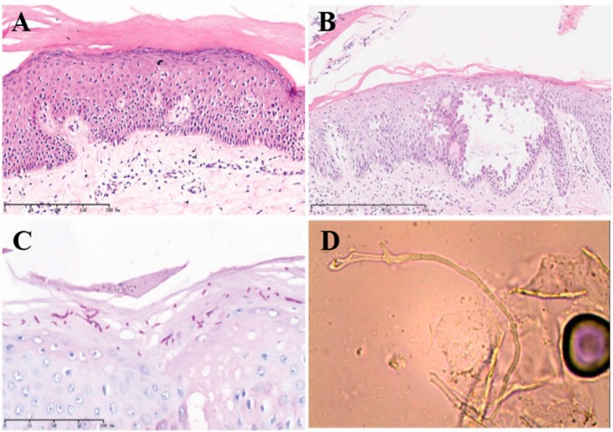
Figure 2.
Histopathological findings of biopsy specimens. (A,B) H and E sections (10X, 40X) of the scalp biopsy showed intraepidermal vesicle formation with acantholytic keratinocytes and overlying scale with parakeratosis; (C) Periodic acid-Schiff (PAS) stains highlighted fungal elements; (D) Trichophyton rubrum was confirmed by culture. Scale bar of A, B and C are in the figures with 200, 800 and 100 µm, respectively.

