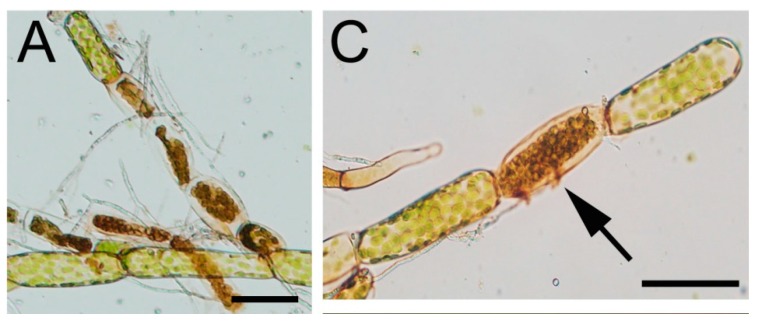
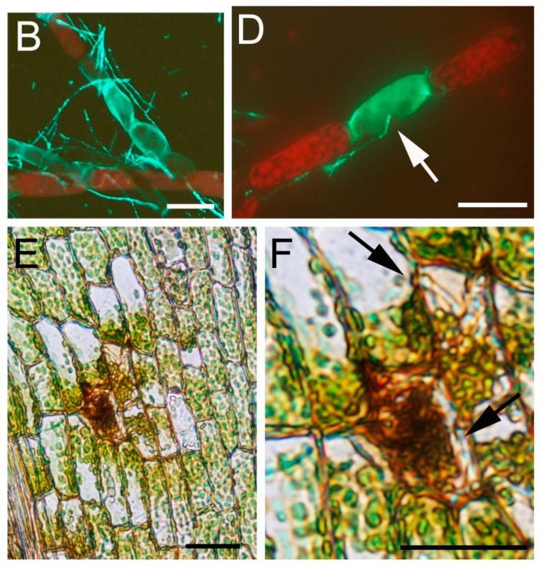
Figure 4.
Cytoplasmic shrinkage and intracellular relocation of chloroplasts after C. gloeo-sporioides infection. (A,C) Protonemal tissues inoculated with C. gloeosporioides; (B,D) Protonemal tissues inoculated with C. gloeosporioides and stained with solophenyl; (E) C. gloeosporioides infected leaf showing chloroplast relocation (F) Closer view of E, hyphae are indicated with black arrows. Arrows in C and D indicate a hypha in contact with a protonemal cell. Scale bars represent 20 μm in A–D and 50 μm in E–F.


