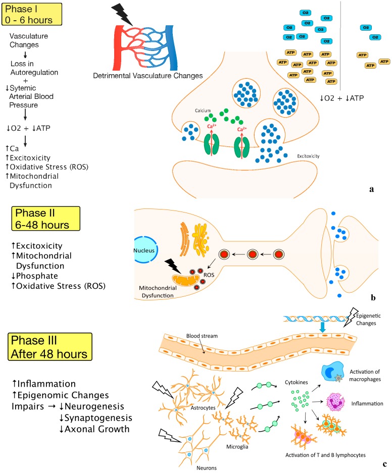
Figure 2.
(a) Vasculature Changes and Primary Energy Failure (Phase I) Legend: A visual representation of the first phase of HIE. Detrimental changes to the vasculature following an HIE insult lead to loss of autoregulation and severe lowering of the systemic arterial blood pressure. This causes a decrease in oxygen, depletion of ATP, as well as increases in excitotoxicity, intracellular calcium, oxidative stress, and mitochondrial dysfunction; (b) Secondary Energy Failure (Phase II). Legend: A schematic representation of the second phase of HIE reveals continued excitotoxicity, oxidative stress, and mitochondrial dysfunction; (c) Chronic Inflammation (Phase III). A pictorial representation of the third phase of HIE shows injury to microglia, neurons, and astrocytes leads to continuous release of cytokines and other detrimental factors causing chronic inflammation which in turn leads to epigenetic changes, as well as impairments of synaptogenesis, axonal growth, and neurogenesis.

