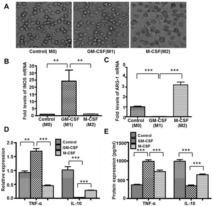
Figure 1.
Macrophage polarization was induced successfully in vitro. Macrophages’ morphology was observed by light microscope, (M0) control macrophages (untreated macrophages); (M1) macrophages stimulated with GM-CSF; (M2) macrophages stimulated with M-CSF (A). Original magnification ×400. INOS (B), ARG-1(C), IL-10 and TNF-α (D) mRNA expression were detected by qRT-PCR. IL-10 and TNF-α (E) protein expression were detected by ELISA. n = 4, ** p < 0.01, *** p < 0.001. M0: Macrophages with no treatment; M1: Macrophages treated with GM-CSF; M2: Macrophages treated with M-CSF.

